


A few weeks ago on April 1, at twenty-one-and-a-half weeks, I had a fetal echocardiogram done. The fetal heart can be examined by ultrasound, similar to an adult echocardiogram, and it is done not by obstetricians, but by pediatric cardiologists. Dr. Dave Atkinson, who was my attending when I was a pediatric resident, and Ning Qi, the echocardiographer, do all the fetal echocardiograms at the hospital where I work. Fetal echos are done when the obstetrician suspects that there might be something wrong with the heart, or there is a family history of congenital heart defects. In my case, Dr. Atkinson and Ning just wanted to look at the twins’ hearts for fun. So I just showed up to the ultrasound room on a Friday afternoon to get another look at the twins on ultrasound. I also brought a videotape so that they could record the images. It’s taken us a little while to transfer the video onto the computer and edit it into four short clips. The first three clips are completely silent. Only the fourth clip has sound.
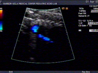
This is an ultrasound video of the aorta of “Twin A”. (By the way, twins are named by their relative position in the uterus. The twin that is lower down in the pelvis is “A”, signifying that she will be delivered first in a vaginal delivery. For me, Twin A is on the right side of my uterus.) The aorta is examined mainly for its overall size and development. There are congenital heart defects where the aorta doesn’t grow well, and either is shortened or narrowed in a specific region. The blue flashes of color represents the blood flow in the aorta, and the fact that it makes a nice candy-cane shape and that it is consistently blue throughout its length means that it is a normally developed aorta.
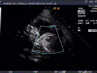
This is a video of the head and torso of Twin A. You can see the face on the left side of the screen and the chest in the middle. The moving dark object in the center of the chest is the heart. The heart itself is examined for abnormalities in structure. Congenital defects of the heart can include underdevelopment of any of the four chambers of the heart, or holes in the walls between chambers. It’s difficult to tell on this image unless you are an experienced reader of echocardiograms but the four chambers of the heart (two atria and two ventricles) appear normal, and there are no holes between chambers, except the one that’s supposed to be there, the foramen ovale.
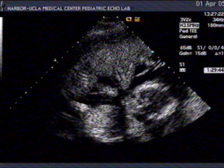
This is a video of both fetuses moving. Twin A is on top Twin B is on the bottom. Twin A is lying with her head on the left side of the image facing down, and you can see her doing a lot of kicking. Twin B is lying with her head on the right side of the image and you can’t really see her legs because they’re bent off to the side and beyond the two-dimensional plane of this image. Between the twins is a thin white line which is the membrane that separates them.
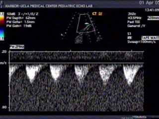
This is a short Doppler ultrasound of the umbilical cord. The umbilical cord consists of two arteries and one vein, and you can aim the probe towards the cord to get an idea of the blood flow going to and from the fetus. The information obtained from one of the umbilical arteries is represented by the waves in the video. As the blood flows from the heart, it makes a pulsating sound which also corresponds to the waveforms. In this quick two second clip, you hear five “whooshes”, which, if you multiply that times thirty, you can calculate that the heart is beating 150 times a minute. As you may know, 150 is slightly fast for a fetal heartbeat, but this is due to the fact that I was about to pass out on the table. By this point in the ultrasound, I had been lying on my back for more than 30 minutes, and I wasn’t getting good blood flow to my own heart (and brain), so I started to feel faint. It’s been well documented that if the mother is uncomfortable, the fetal heart rate goes up. When I shifted to lying on my side, I felt much better, and the fetal heart rate went back to normal (around 130 beats per minute).